

Are you tired of overcomplicated stereology applications? Would you like to speed up volume estimation from a set of histology sections? With microDimensions Outspace™ you are guided by an intuitive wizard to quantify volumes based on the Cavalieri estimator. Save up to 30% time in your studies due to increased automation compared to conventional stereology software!
With microDimensions Outspace you can significantly speed up your lab work – let the software automatically detect and align all sections on your glass slides instead of struggling with rotating or flipping them manually. Selection of specific sections, definition of regions of interest, and volume estimation is all bundled in microDimensions Outspace: a solution that can be seamlessly integrated into your workflow, from the available image set to the final result export.
For a chance to win a free license for microDimensions Outspace worth USD 4,500,
register now for the raffle! *
Get Outspace now to save clicks and time with computer-assisted stereology.
* For Research Use Only. Not for use in diagnostic procedures. Entries are collected during the entry period from June 15, 2015, 0:01am EDT to July 31, 2015, 11:59pm PDT. An entry requires the submission of the full name, e-mail address, telephone number, organization, country, and answering up to five short questions using the form provided at micro-dimensions.com/events/2015/6/15/outspace-raffle. Only one submission per person is allowed. Employees, business partners, or competitors of microDimensions are not allowed to participate. Among all entries one winner will be determined by random draw. The winner will be notified by e-mail and will receive a license for microDimensions Outspace (subject to the end user license agreement available at micro-dimensions.com/eula) that is valid from September 1, 2015 to February 29, 2016 (corresponding to a value of USD 4,500) at no cost and with no automatic extension or any other obligations. The prize cannot be claimed in cash.
![Reconstructed brain region showing complex interactions between blood vessels (red, CD31), cancer cells (green, GFP), and astrocytes (white, GFAP)[Image courtesy of Sho Fujisawa, Memorial Sloan Kettering Cancer Center]](https://images.squarespace-cdn.com/content/v1/54d89682e4b0a80ddb2c75f2/1429773382082-WE49O686ATGXXH9YWCTP/Reconstructed+brain+region)
![Reconstructed CD31-positive blood vessel (red) and spot detection of pMAPK-positive cells with color-coded distance information[Image courtesy of Sho Fujisawa, Memorial Sloan Kettering Cancer Center]](https://images.squarespace-cdn.com/content/v1/54d89682e4b0a80ddb2c75f2/1429773780111-7W0KWS77YQN25ERHYGQZ/Reconstructed+blood+vessel)
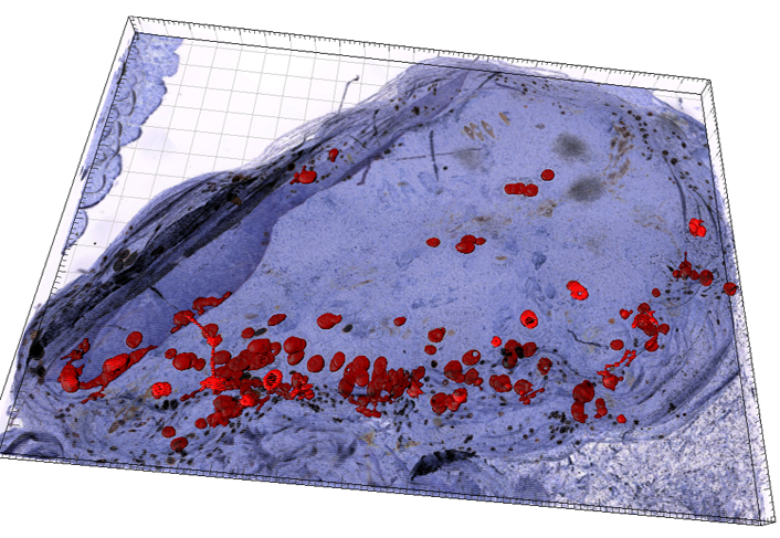
![CD31 stained blood vessels in reconstructed myxofibrosarcoma[Image courtesy of Sho Fujisawa, Memorial Sloan Kettering Cancer Center]](https://images.squarespace-cdn.com/content/v1/54d89682e4b0a80ddb2c75f2/1429773071949-TLR62EEEAFIGQQ68NZES/Blood+vessels+in+reconstructed+myxofibrosarcoma)


