

Apart from all the technical differences in image acquisition brightfield and fluorescence images can provide pathologists with different insights on the assay. While brightfield images (e.g. stained in H&E) are the primary choice for observing the morphology of the tissue, fluorescence images are better for the visualization of the cellular details. Digital multiplexing of brightfield and fluorescence images can combine the benefits of both worlds while maintaining the individual characteristics of each of the modalities. Many digital pathology scanners available today are equipped to scan images in both brightfield and fluorescence mode. When using both modes for a tissue sample, adjoined sections are typically prepared and scanned separately, so the resulting whole slide images (WSIs) need to be reviewed or analyzed individually. By using image registration the images can be co-located to facilitate visual evaluation or automated image analysis.
Overview Brightfield vs. Fluorescence Whole Slide Imaging
Brightfield
Fluorescence
Image Characteristics
Shows the whole structure of the tissue incl. surroundings
Shows only what has been stained positively, better at specific cellular location
Ease of Reading
Reading slides requires years of histopathology expertise
Easier to read due to eliminated background
Image Acquisition
Faster and easier, equipment is often less costly
More complex setup, can take longer and be more expensive
Image Channels
Images are acquired and stored in RGB mode
Images are acquired and stored with individual channels
Common Staining
H&E, IHC, CISH, other visible stains
Immunofluorescence, FISH
Typical Usage
Most commonly used in clinical routine due to ease of use, FDA clearance for various vendors
Used in research applications, FDA clearance and widely used in clinical routine on FISH assays
How to Combine the Best of Both Worlds
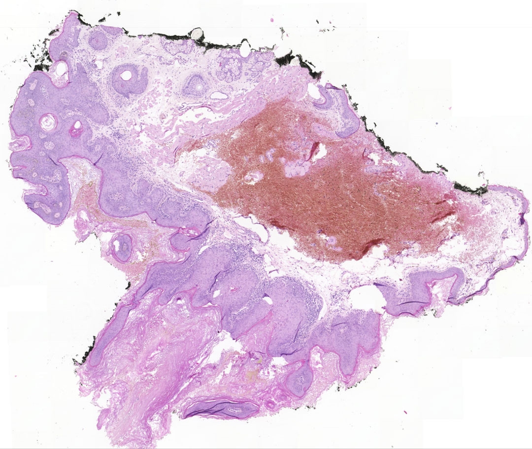
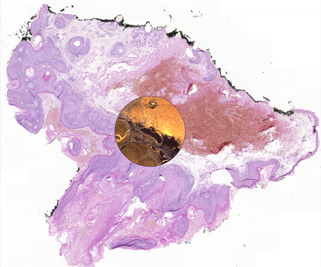
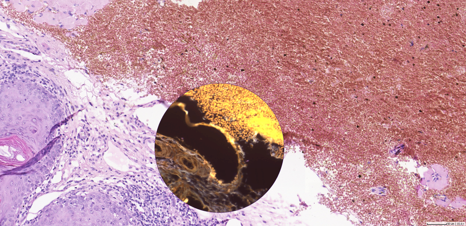
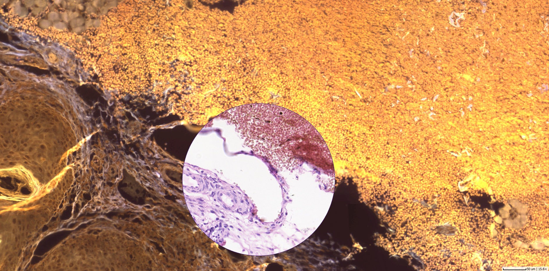
Aligned images in brightfield (H&E) and fluorescence (Rhodamine, DAPI, FITC, Cy5)
Aligning images from multiple modalities can help aggregating information from consecutive sections stained in histochemical and fluorescent dyes, e.g. protein expression and gene amplification by aligning immunohistochemical (IHC) markers with fluorescence in-situ hybridization (FISH).
The aligned images can then be used to facilitate the visual evaluation by a pathologist or automated image analysis. There is a wide range of applications conceivable for this method from cancer research and diagnosis to drug development and biomarker discovery. One of the most prominent ones might be research and diagnosis of HER2-positive breast cancer.
Sample Application: Multiplex-Analysis of HER2-Positive Breast Cancer
HER2-positive breast cancer tends to be more aggressive, so targeted therapies have been developed for the treatment of this particular type of cancer. Existing image-based solutions carry out the pathological scoring in breast cancer either with a scoring based on immunohistochemical IHC staining to mark the HER2 protein on the cell membrane, or a scoring based on FISH to mark the HER2 ribonucleic acid (RNA) that contains the genetic information to produce the HER2 protein. By using the aligned images the scoring of HER2 protein overexpression and gene amplification could be combined. This can be achieved by employing automatic image registration to align consecutive cuts that have been stained with IHC HER2 and FISH. After the alignment, similar regions on the sections can be identified quickly and an analysis of the results can be related on a modal and spatial correlation. This could help to overcome the limitations of current tests and help reduce uncertainty in diagnosis.
BRIGHTFIELD and Fluorescence Multiplexing as Part of Slidematch

microDimensions will introduce the capability to align brightfield and fluorescence whole slide images in the next release of Slidematch - a software for the alignment of differently stained sections. With the new feature you can align WSIs in both modalities at a very high precision only limited by the scanner resolution. The import wizard allows to load images in formats from different vendors and in different resolutions in the same batch. The images can be stored as an aligned image series in the .svs, .tif, or .ims format for viewing or automated image analysis.
The new feature will also be presented at Pathology Visions 2016, 23 - 25 October in San Diego, California. Come by our booth #310 to learn more. We are looking forward to your feedback!