Experience our New, Faster Versions of Slidematch and Voloom

Comment

Experience our New, Faster Versions of Slidematch and Voloom

BENEFIT FROM A MASSIVE EFFICIENCY GAIN

Fasten your seatbelt: Slidematch, our software for alignment of differently stained sections as well as Voloom, 3D histology software for reconstruction of serial sections, are now available as new, improved versions for evaluation and purchase.

Our development team was able to speed up data processing algorithms by an order of a magnitude. As a result, Slidematch 2.0 saves aligned data up to ten times quicker which will certainly take your research analysis to the next level. In the new Voloom 3.0 the speed of deformable data export was significantly increased making high-resolution image data ready to use in other programs faster than ever. 

Check out our product pages to find out more about benefits and fields of application of our software, to request trial licenses or a quote:


Comment

microDimensions achieves important milestone with focus on Digital Pathology

Comment

microDimensions achieves important milestone with focus on Digital Pathology

2017 was a very successful year for microDimensions: its revenue had more than doubled and the team overachieved the ambitious goals set. This is an important milestone for the young company dedicated to Digital Pathology. microDimensions’ growth goes hand in hand with the expanding digital transformation in healthcare, the latest advances in image processing technology as well as the disruption of medical research and diagnostics by computational pathology and artificial intelligence.

The motivated team of computer scientists, business and marketing specialists is looking forward to achieving the next strategic milestone in 2018 by positioning their platform anyslide.com as the only hub for cross-referenced digital pathology slides (#digitalpathologyhub).

2018-02-26-Corp-News---Milestone---Webpage.png

“With this new platform, microDimensions is committed to serve customers such as Life Sciences companies looking for high quality curated data to train their Artificial Intelligence algorithms in pathology or to publish their scientific results to the medical community (#PrecisionMedicine).” says Ralph Gloser, newly appointed Chief Commercial Officer. “Public and private institutions have also shown their interest in using Anyslide to effectively train their students and provide modern tools of education through digitization and online collaboration.”

Further information on Anyslide can be found on anyslide.com. Visitors of the USCAP 2018 in Vancouver in March can experience the platform live at booth #543 and discuss how it helps to foster knowledge exchange and to accelerate medical progress.

Comment

New image sharing platform Anyslide opens up digital pathology to everyone

Comment

New image sharing platform Anyslide opens up digital pathology to everyone

microDimensions launches new cloud-based image sharing and collaboration platform, helping to overcome some of the major hurdles on the way to digital pathology.  

Digital Pathology is happening. While the advantages are evident and experts already predict a new generation of pathologists, only few laboratories and institutes have started planning and investing in fully digital work-flows. At the same time, the demand for professional online exchange and a convenient way of sharing educational or rare cases is growing with the aim to support a more precise and profound health care.

To enable pathologists to start working digital with their existing set-up today, microDimensions is now launching AnyslideTM, a new cloud-based digital pathology platform. Pathologists can easily sign up for free, share and collaborate on digital histology images in seconds using their desktop browser, tablet or smartphone. By eliminating the need for a major investment or effort put into installation and integration, Anyslide helps organizations to overcome many of the major hurdles on the way to digital pathology.

“We have designed Anyslide as a global pathology hub for the needs of today’s pathologists - no matter how advanced their labs are on the digital journey, and no matter what devices they use to interact online on images” says Dr. Martin Groher, CEO of microDimensions.

Anyslide is driven by pathologists' work-flows in education, research and professional exchange, and is dedicated to reducing mouse clicks for the most common tasks:

  • Online collaboration with a colleague or in groups: Case images can be shared and discussed privately by granting access to specific users and letting them comment on conclusions.
  • Public sharing: Pathologists can upload, describe and annotate exciting findings and share them with their social network, e.g. on Twitter.

Anyslide supports the upload of images in various formats (JPEG, PNG, TIFF, GIF, and Whole Slide Images*) as well as live capture from microscope cameras. The platform provides simple tools to edit and annotate, comment on, and share images in one unified application. Signing up and using Anyslide is free of charge.   

“As daily routines of pathologists and the technologies around them are evolving constantly, we are planning to let Anyslide evolve too - from the support of whole slide images to the integration of further social networks and advanced group collaboration functionality.” continues Groher. “But also services based on image processing and machine learning algorithms are possible, where big data approaches could open up huge potential for automatic classification and detection tools as well as scoring aids.”

To find out more about Anyslide and to sign-up today, please visit https://into.anyslide.com.

Anyslide will be presented at booth #36 at the 29th European Congress of Pathology (ECP) in Amsterdam, 2nd - 6th September 2017. To schedule an appointment, please contact anyslide@micro-dimensions.com.


* Whole Slide Image (WSI) support will be available in the Anyslide Professional plan

Google is a registered trademark of Google, Inc. in the United States and other countries. Twitter is a registered trademark of Twitter, Inc. in the United States and other countries.


Comment

Slidematch™ now features Alignment of Brightfield and Fluorescence Whole Slide Images

Comment

Slidematch™ now features Alignment of Brightfield and Fluorescence Whole Slide Images

microDimensions launches a new version of its high precision whole slide alignment software Slidematch. With the new version users can align digital whole slide images from consecutive sections in brightfield and fluorescence mode, e.g. for the evaluation of the HER2 status of cancer patients.  

While brightfield images (e.g. stained in H&E) are the primary choice for observing the morphology of the tissue, fluorescence images are better for the visualization of the cellular characteristics. Digital multiplexing of brightfield and fluorescence images can combine the benefits of both worlds while maintaining the individual characteristics of each of the modalities. Many digital pathology scanners available today are equipped to scan images in both brightfield and fluorescence mode. When using both modes for a tissue sample, adjoined sections are typically prepared and scanned separately and the resulting whole slide images (WSIs) need to be reviewed or analyzed individually. Aligning images from multiple modalities can help aggregating information from consecutive sections stained in histochemical and fluorescent dyes, e.g. protein expression and gene amplification by aligning immunohistochemical (IHC) markers with fluorescence in-situ hybridization (FISH). The aligned images can then be used to facilitate the visual evaluation by a pathologist or automated image analysis.

An application example is the evaluation of the HER2 status of breast cancer patients. Typically the pathological scoring of the HER2 status  in breast cancer is either based on immunohistochemical IHC staining to mark the HER2 protein on the cell membrane, or on FISH to mark the HER2 ribonucleic acid (RNA) that contains the genetic information to produce the HER2 protein. By using the aligned images the scoring of HER2 protein overexpression and gene amplification could be combined to achieve a higher precision.

There is a wide range of applications conceivable for this method from cancer research and diagnosis to drug development and biomarker discovery.

Slidematch can perform the alignment of the digital slides at a very high precision only limited by the scanner resolution. The import wizard allows to load images in formats from different vendors and in different resolutions in the same batch. The new version now also supports the import of images in proprietary formats from Roche-Ventana, Olympus and PerkinElmer. The images can be stored as an aligned image series in the .svs, .tif, or .ims format for viewing or automated image analysis. More Infomation is available at http://micro-dimensions.com/slidematch.

Slidematch will be presented at booth #627 at Molecular Medicine Tri-Con 2017,  20 - 22 February in San Francisco. To schedule an appointment at one of these exhibitions, please send an email with your preferred date and time to sales@micro-dimensions.com.

Comment

Comment

Upcoming Digital Pathology Webinars: 3D Histology and Multiplex Analysis of Brightfield Images

microDimensions is hosting two compact digital pathology webinars back to back on Oct 11th. The first session on "3D Histology" will show how to reconstruct 3D volumes from serial histological sections (brightfield or fluorescence), how to view and analyze them and how to perform volume measurements e.g. of tumors.  The second session on "Multiplex Analysis of Brightfield Images" will focus on the automated alignment of differently stained brightfield images and how the aligned images can be used to speed up and facilitate the following analysis. Both webinars will be presented by Andreas Keil (PhD),  the VP of Business Development at microDimensions. 

Dates: 

  • "3D Histology", Tuesday, 11 Oct. 2016, 8 am PDT / 11 pm EDT / 17:00 CEST
  • "Multiplex Analysis of Brightfield Images", Tuesday, 11 Oct. 2016, 8:30 am PDT / 11:30 pm EDT / 17:30 CEST

The attendance is free of charge. Interested attendees can register here for one or both sessions here.


Meet us at these upcoming exhibitions:

  • Pathology Visions, 23 - 25 October 2016, San Diego, U.S.A.
  • Munich Biomarker Conference, 29 - 30 November, Munich, Germany
  • Digital Pathology Congress Europe, 1-2 December 2016, London, UK

To schedule an appointment at one of these exhibitions, please send an email with your preferred date and time to sales@micro-dimensions.com.

Comment

microDimensions receives Series A Financing for Digital Pathology Solutions

Comment

microDimensions receives Series A Financing for Digital Pathology Solutions

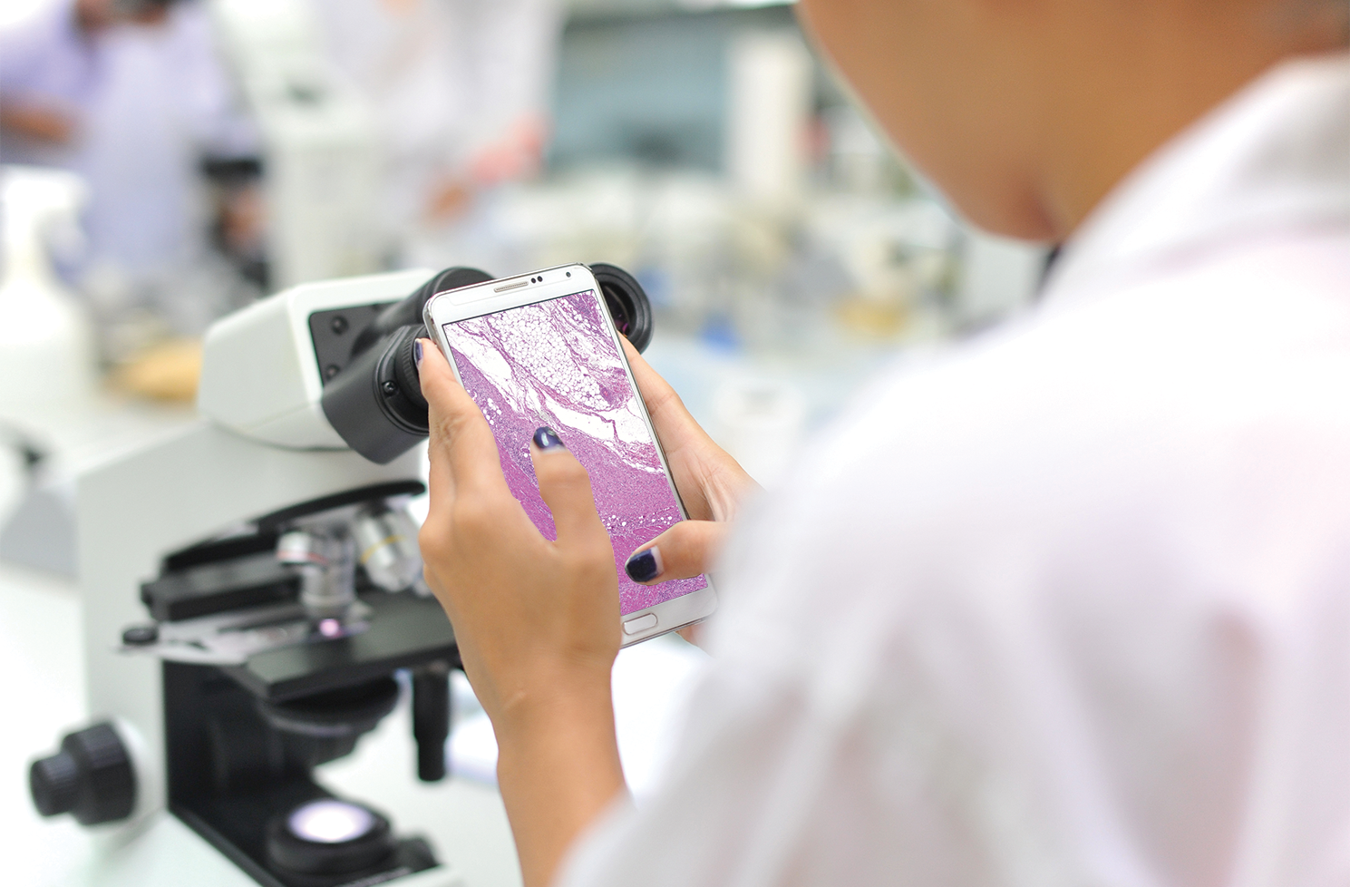
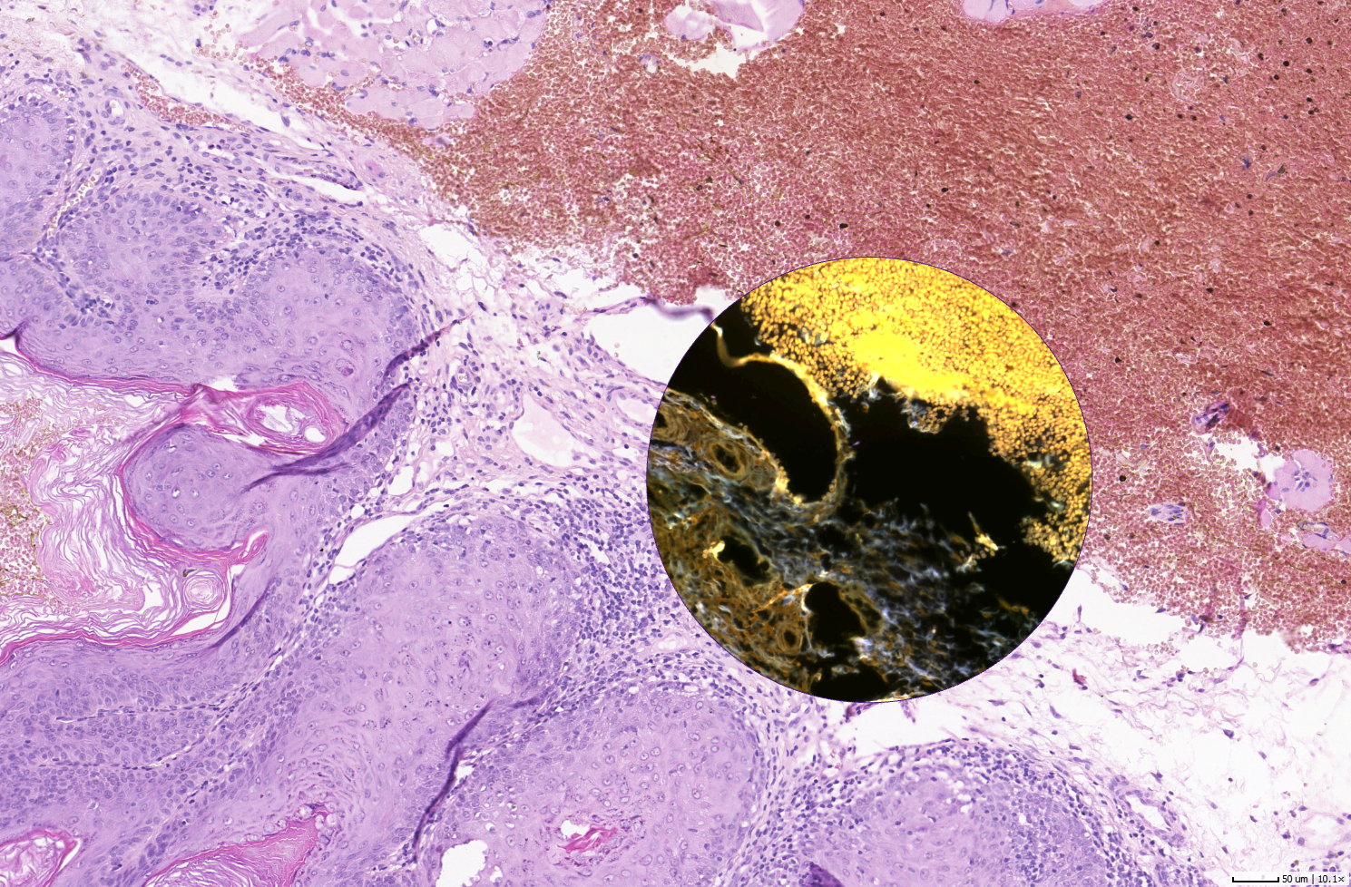
microDimensions GmbH, a Munich-based software and service provider for digital pathology that specializes in automated image analysis raised a Series A financing round. The company’s unique image processing technology can significantly speed up and automate workflows in (pre-)clinical drug development studies and clinical diagnosis. A consortium of five investors, led by Business Angel Dr. Adriaan Hart de Ruijter and complemented by High-Tech Gründerfonds, Bayern Kapital and a group of business angels, provided a seven-digit investment to enable microDimensions to further extend its product portfolio. 

The market for digital pathology is estimated to reach a market volume of 5.2 Billion USD up until 2020. “The main drivers for digitization are the stagnating number of pathologists and the high complexity of biomarker analysis. This increases the demand for efficient workflows and cost-effective solutions in the life science industry as well as clinical pathology institutions.” explains Dr. Martin Groher, CEO of microDimensions. “The fresh capital enables us to fully exploit the potential of state of the art technologies such as automated image analysis, big data and deep learning to develop software solutions that help our customers to find the right answers to major health threats such as cancer.” 

microDimensions has successfully launched a series of software products that are used by major pharma, biotech, and medtech players, and various university pathology departments. The Series A funding will be used to further develop the product line for biomarker image analysis. Besides its standardized software solutions, microDimensions offers the development of custom applications as well as a range of image analysis services. “Many research organizations are pioneers in their field and their software requirements cannot be fulfilled with an off-the-shelf product. Based on the technology and in-house expertise microDimensions can provide them with tailored solutions and on demand image analysis services in exactly the way they need it.” says Dr. Adriaan Hart de Ruijter.

About microDimensions
microDimensions develops and distributes software for microscopic image processing and analysis. Their solutions and services can be tailored to individual requirements and seamlessly integrated into digital pathology workflows.  microDimensions’ cutting-edge products Voloom®, Slidematch™, and Zoom are the world’s fastest tools for convenient and accurate 3D histology reconstruction, whole slide image alignment, stereology, and digital pathology viewing. They enable pharmaceutical and biotech companies to accelerate early drug testing and allow clinical and research organizations to gain new insights into cancer, multiple sclerosis, chronic infections, and other diseases.

Contact:
microDimensions GmbH
Dr. Martin Groher
Rupert-Mayer-Str. 44 // 64.07
81379 München
Germany
Tel.: +49.89.1894253.30
info@micro-dimensions.com
www.micro-dimensions.com

About Bayern Kapital
Bayern Kapital GmbH, based in Landshut, was founded on the initiative of the Bavarian government in 1995. It is a wholly-owned subsidiary of the Bavarian LfA Förderbank. As the venture capital organisation of the Land of Bavaria, Bayern Kapital provides equity capital financing for the founders of young innovative technology companies in Bavaria.

Presently Bayern Kapital manages eleven investment funds with a total volume of around €340m. So far, it has invested almost €227m in 245 innovative companies in the fields of technology in various sectors including life science, software & IT, medical technology, materials and new materials, nanotechnology and environmental technology.

In this way, almost 5000 long-term jobs in sustainable companies have been created in Bavaria.

Contact:
IRA WÜLFING KOMMUNIKATION GmbH
Dr. Reinhard Saller
Tel.: +49. 89. 2000 30-30
bayernkapital@wuelfing-kommunikation.de

About High-Tech Gründerfonds
High-Tech Gründerfonds invests in young, high potential high-tech start-ups. The seed financing provided is designed to enable start-ups to take an idea through prototyping and to market launch. Typically, High-Tech Gründerfonds invests EUR 600,000 in the seed stage, with the potential for up to a total of EUR 2 million per portfolio company in follow-on financing. Investors in this public/private partnership include the Federal Ministry of Economics and Energy, the KfW Banking Group, as well as strategic corporate investors including ALTANA, BASF, Bayer, B. Braun, Robert Bosch, CEWE, Daimler, Deutsche Post DHL, Deutsche Telekom, Evonik, Lanxess, media + more venture Beteiligungs GmbH & Co. KG, METRO, Qiagen, RWE Innogy, SAP, Tengelmann and Carl Zeiss. High-Tech Gründerfonds has about EUR 576 million under management in two funds (EUR 272 million HTGF I, EUR 304 million HTGF II).

Contact:
High-Tech Gründerfonds Management GmbH
Dr. Marianne Mertens
Schlegelstr. 2
53113 Bonn
Tel.: +49 228 823001-00
Fax: +49 228 823000-50
m.mertens@htgf.de
www.high-tech-gruenderfonds.de

Comment

Comment

New Business Development Manager: Irene Meliciani Joins the microDimensions Team

microDimensions is pleased to announce the appointment of Irene Meliciani as Business Development Manager. Irene has graduated in genomics biotechnology from La Sapienza University in Rome and holds a PhD in computational biochemistry obtained from Karlsruhe Institute of Technology. In addition to her scientific qualification she has a developed strong background in business development and sales during her professional experience in the pharma and biotechnology industry since 2012.  Having lived in Belgium, Italy, Germany and Spain, Irene is a true European and supports microDimensions’ international business with her wide range of language skills. As one of her main tasks, Irene will be in charge of developing the growing image analysis service business. Based on its in-house software development capabilities, microDimensions is able to provide a range of services that can be tailored to customer requirements, if needed.

“We are very pleased to extend our team with an experienced and passionate professional like Irene.” says Marco Feuerstein, Managing Director.

The microDimensions team has grown from 7 to 10 employees since the beginning of this year. The Munich based software company is planning to further extend their staff with one more Business Developer and one Software Developer during the next months. Open positions can be found at micro-dimensions.com/careers.


Meet us at these upcoming exhibitions:

Digital Pathology Congress USA, 14-15 July 2016, Philadelphia, USA
Digital Pathology Congress Europe, 1-2 December 2016, London, UK

To schedule an appointment at one of these exhibitions, please send an email with your preferred date and time to sales@micro-dimensions.com.

Comment

Zoom 2.0: Digital Pathology Viewer Now Features Multiple Image Viewing and Annotations

Comment

Zoom 2.0: Digital Pathology Viewer Now Features Multiple Image Viewing and Annotations

microDimensions has launched an upgraded version of its free digital pathology viewer Zoom, including a set of features to facilitate collaboration of researchers working on digital whole slide images. 

The version 2.0 enables loading and viewing of a series of previously aligned images such as 3D reconstructions or aligned images with different staining that have been created with other software products such as Slidematch™. With the new peephole function two aligned images can be viewed at the same time, e.g. to compare the response of tumors to different biomarkers.

The new annotations functionality offers the possibility to mark areas of interest, add comments and share these with others. For easy editing and comparison of areas of interest annotations can be copied to the other images of a series or volume. Annotations can also be imported from and exported to Aperio ImageScope. 

“With the new version of Zoom we have created an easy to use but yet powerful tool for image viewing and collaboration between researchers, that is available for free to anyone.” says Dr. Marco Feuerstein, COO of microDimensions.

Zoom enables fast viewing of whole slide images from all major hardware vendors as well as 3D reconstructions generated with Voloom. The version 2.0 also supports pyramidal and 3D TIFF files. The intuitive channel settings allow simple adjustment of rendering settings (brightness, contrast, gamma) and turning on/off of individual channels for an unlimited number of channels of 8 bit and 16 bit data, no matter whether they were acquired by brightfield, electron microscope, fluorescence, or laser scanning systems. 

The software is available free of charge at micro-dimensions.com/zoom. For more information, please watch our video tutorial


Meet us at these upcoming exhibitions:

European Congress on Digital Pathology, 25-28 May 2016, Berlin, Germany
Digital Pathology Congress USA, 14-15 July 2016, Philadelphia, USA
Digital Pathology Congress Europe, 1-2 December 2016, London, UK

To schedule an appointment at one of these exhibitions, please send an email with your preferred date and time to sales@micro-dimensions.com.

Comment

Comment

US Patent Granted for Fast Image Registration Technology

microDimensions announced today that the United States Patent and Trademark Office has issued the US Patent 2015/0016703 A1 for its method for image registration of sections, in particular of histological sections. The patented technology enables fast and accurate alignment of whole slide and other large images up to the scanned resolution.

A single histological section can easily require more than 1 GB of storage space. When handling multiple images of this size during the alignment the limits of storage capacity available today can rapidly be exceeded. To solve this problem and enable efficient digital pathology workflows microDimensions has developed a unique image registration technology that uses a quadtree-like data structure to partition an image into cells with individual local transformations on multiple resolution levels. This can tremendously speed up the alignment of a region of interest at high resolution, as only a subset of cells is to be processed. The use of local transformations significantly increases the final alignment quality to an accuracy that is only limited by the scanner resolution.

The patented technology is used in the 3D histology software Voloom® to enable fast 3D reconstructions from serial sections. Voloom can reconstruct 3D volumes in a matter of minutes and by this enables researchers to effectively use 3D reconstructions in their daily workflows. More information on Voloom

Slidematch™, the latest addition to the microDimensions product line, uses the registration technology for the alignment of differently stained sections to perform e.g. multiplex analysis or compute scores for improved cancer classification. More information on Slidematch

microDimensions is a spin-off company of the Technische Universität München and has a strong scientific background in the field of image registration. The patented technology was developed in house. “The patent is an important milestone for the company and a major component of our intellectual property. Fast image registration is essential to our products, since it enables a seamless combination of large image files as required by digital pathology workflows.” says Martin Groher, CEO of microDimensions.

Comment

Comment

New Tutorial Series for 3D Histology Software Voloom

3D Reconstruction of the Colon of a Mouse Embryo

Together with the new version of its 3D histology software Voloom microDimensions is launching a complete updated online tutorial series. The tutorials are aimed at pathologists, researchers and scientist interested in 3D reconstruction and analysis of digital whole slide images with Voloom and do not require any previous knowledge. The different chapters cover the reconstruction process, usage, segmentation & analysis, color deconvolution, and export of data in short units of two to seven minutes.

The latest version (2.6) of Voloom includes improvements concerning the interoperability with third party products such as extended support of CZI (Zeiss) image pyramids, exporting SVS/AFI files (Leica/Aperio) or loading of IMS file series (Bitplane) as 3D volumes. Voloom 2.6 also supports network licences for easy operation of the software on multiple workstations. 

“Even if the latest updates in Voloom affect the software more on a technical level, it will improve the overall user experience by ensuring a smooth operation within the customer’s infrastructure.” says Dr. Hauke Heibel, CTO of microDimensions.

Both the tutorial series as well as a free trial version of Voloom are available on micro-dimensions.com/voloom


Meet us at these upcoming exhibitions:

  • European Congress on Digital Pathology, 25-28 May 2016, Berlin, Germany
  • Digital Pathology Congress USA, 14-15 July 2016, Philadelphia, USA
  • Digital Pathology Congress Europe, 1-2 December 2016, London, UK

To schedule an appointment at one of these exhibitions, please send an email with your preferred date and time to sales@micro-dimensions.com.

Comment

microDimensions Slidematch Enables Multiplexed Protein Measurement on Consecutive Sections

Comment

microDimensions Slidematch Enables Multiplexed Protein Measurement on Consecutive Sections

microDimensions, software and service provider for digital pathology image analysis solutions, recently launched Slidematch™, the one-of-a-kind software for the alignment of differently stained brightfield sections in full resolution. microDimensions Slidematch loads a series of consecutive whole slide images, aligns them using patent-pending registration technology, and saves them as another series of aligned whole slide images. The whole alignment process requires a minimum of user interaction, so study workflows including e.g. multiplexed protein measurement can be sped up significantly.

“Compared to other software for co-registration, the accuracy and robustness of microDimensions’ alignment technology is unmatched, especially when we move on a high magnification level”, says Stefan Hamann, senior scientist in Research Technology at Biogen USA. “Due to the high level of automation of Slidematch we can significantly speed up our studies and create more significant readouts.”

Amongst others, aligned whole slides created with Slidematch can be used for:

  • Transferring marked regions of interest from one section to other sections
    For example tumor regions and hotspots can be marked in e.g. H&E, PCK, or organelle-stained sections and transferred to IHC sections
  • Multiplexing of protein expression
    Signals coming from aligned IHC sections with different staining can easily be correlated to measure the abundance of many proteins
  • Correlation of protein expression and gene amplification
    Diagnosis can be improved comparing the spatial relation of e.g. HER2 IHC and ISH sections

This all leads to extreme speed-ups in analysis tasks such as score computation. For instance, after tumor hotspots are marked in an H&E section, they can be automatically transferred to CD3 and CD8 IHC sections, so the ratio of cytotoxic and normal T cells can be computed.

For more information, including a link to request a free trial version, please visit the Slidematch product page micro-dimensions.com/slidematch.


Meet us at these upcoming exhibitions:

  • European Congress on Digital Pathology, 25-28 May 2016, Berlin, Germany
  • Digital Pathology Congress USA, 14-15 July 2016, Philadelphia, USA
  • Digital Pathology Congress Europe, 1-2 December 2016, London, UK

To schedule an appointment at one of these exhibitions, please send an email with your preferred date and time to sales@micro-dimensions.com.

Comment

Comment

Voloom 2.5: Full-Resolution Whole Volume Export

microDimensions today released an update to Voloom, its world leading 3D histology software for the fast reconstruction of serial sections. microDimensions Voloom 2.5 now provides deformable full-resolution export of whole reconstructed volumes. This makes it easy to further process and analyze reconstructed volumes with other solutions in the same resolution as the original whole slide images. Full-resolution volumes can be stored as Bitplane Imaris .ims files, with more export formats to come in the future. Alignments can also be pre-computed up to the full resolution during the reconstruction process, so that subsequent visualization, analysis, and export is sped up significantly.

Moreover, version 2.5

  • is compatible with OpenGL 3.3 capable Intel graphics boards,
  • implements a new intelligent section ordering algorithm,
  • improves the MDV format and loading times for fully registered data sets, and
  • adds support for non-standard DPI settings and retina displays.

For more information please visit the Voloom product page micro-dimensions.com/voloom.

We sincerely invite you to register for one of our upcoming free webinars on volumetric histology in order to get an insight into the features and capabilities of Voloom. The next demo webinars are scheduled for:

  • Tuesday, 10 November 2015, 9am PST / 12pm EST / 18:00 CET
  • Thursday, 17 December 2015, 8am PST / 11am EST / 17:00 CET
  • Monday, 25 January 2016, 10:00 CET / 18:00 JST

Register now at micro-dimensions.com/webinars.

Comment